


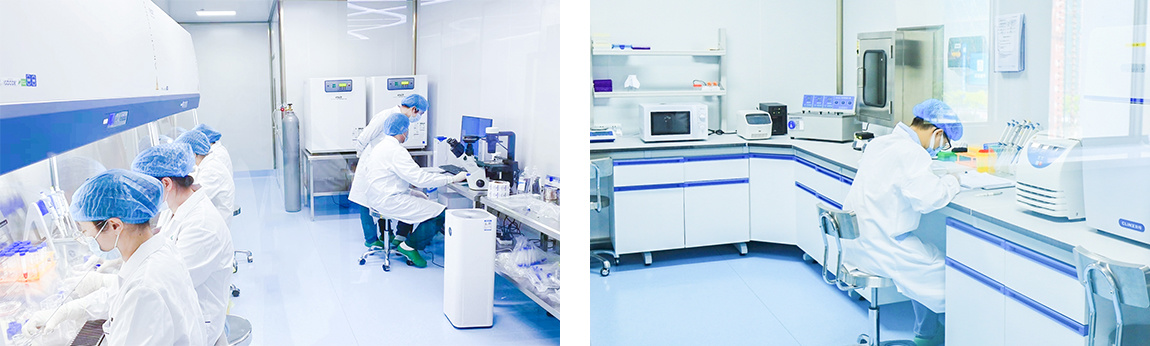
HUAREN MEDICAL TECHNOLOGY
R & D SERVICES
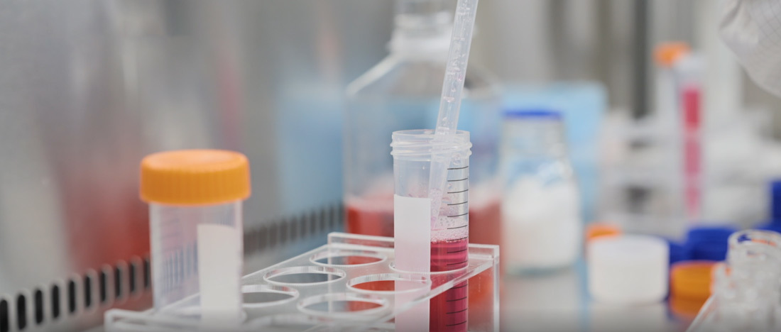
Basic Scientific Research
Basic Scientific Research

Histopathology Experiments
• He Staining Method
Hematoxylin-eosin staining, referred to as HE staining, is the most commonly used staining method.
 |
Hematoxylin (hematoxylin) is a cationic dye that stains basophiles in the nucleus of cells blue-violet. |
 |
Eosin is an anionic dye that stains cytoplasm, collagen fibers, etc. pink. |
A well-made HE section is the key for pathologists to make a correct diagnosis, and HE staining is a routine staining method widely used in domestic and international pathology diagnosis. The quality of the section can directly affect the timely and accurate diagnosis of the disease.
|
• Tunel Ttaining Tunel staining i.e. in situ terminal transferase labeling technique. Apoptotic cell DNA double-strand breaks or a gap in one strand produce a series of 3'-OH ends, and under the action of deoxyribonucleotide terminal transferase (TdT), the derivatives formed by deoxyribonucleotide and biotin are labeled to the 3′ end of the DNA, so as to carry out apoptotic cell detection. TUNLE staining is a combination of molecular biology and morphology. In situ staining of intact single apoptotic nuclei or apoptotic vesicles accurately reflects the most typical biochemical and morphological features of apoptotic cells. • Masson Stain Ponceau S aniline blue staining, is one of the staining methods used for the detection of collagen fibers in animal tissues, able to collagen fibers stained blue, myofibrils, and erythrocytes red, can be used for identification of collagen fibers and myofibrils and to show that a variety of tissues of collagen fibers content and the degree of fibrosis.
|
|
• Sirius Red Stain Both Sirius Scarlet and KWS are strong acidic dyes, which can easily bind to the basic groups in collagen molecules and adsorb firmly. Polarized light microscopy shows that collagen fibers have positive uniaxial birefringent light properties, and the combination with KWS-Tenwolf Scarlet enhances the birefringence and improves the resolution, thus distinguishing the two types of collagen fibers. After the sections of undecalcified bone tissue were stained with Sirius Scarlet, under normal light microscope, collagen fibers were red or bright red, and the others were yellow; under polarized light microscope, collagen fibers of type I were strong orange-yellow or bright red, and collagen fibers of type III were green. • Oil Red O Coloring The staining mechanism of oil red O on lipid droplets is generally considered to be physically solution action or adsorption, which lends solution action to lipid staining, i.e., when oil red O is first dissolved in 60% isopropanol, and then when the section is immersed in the oil red O staining solution, the solubility of oil red O in the tissue lipids is higher than that in 60% isopropanol, and so during staining, the oil red O transfers from the 60% isopropanol into the lipids to make the droplets show red color.
|
|
• safranin O-Fast green Staining In morphological studies involving articular cartilage and subchondral bone, it is often necessary to use a combination of dyes to visualize the histological structure. The safranin O-fast green stain, which originated in the 1960s, is favored for its ability to visualize the structure of articular cartilage, subchondral bone and bone tissue. Cartilage appears red and osteogenic bone appears green. • Immunofluorescence Staining The method of tracing or checking the corresponding antigen with fluorescent antibody is called fluorescent antibody method. The method of tracing or checking the corresponding antibody with known fluorescent antigen marker is called fluorescent antigen method. These two methods are collectively referred to immunofluorescence technology. The fluorescent pigment can not only bind to antibody globulin for detection or localization of various antigens, but also bind to other proteins for detection or localization of antibodies, but in practice the fluorescent antigen technology is rarely applied, so it is customary to refer to as the fluorescent antibody technology, or immunofluorescence technology. The fluorescent antibody method is more commonly used. Immunofluorescence technology to show and check the cell or tissue antigen or semi-antigenic substances and other methods are called immunofluorescence cell (or tissue) chemistry technology.
|
|
• Immunohistochemical Staining Immunohistochemistry is the application of the basic principle of immunology - antigen-antibody reaction, i.e., the principle of specific binding of antigen and antibody, to determine the antigens (peptides and proteins) in tissue cells through the coloration of colorants (fluorescein, enzymes, metal ions, and isotopes) labeled with antibodies through a chemical reaction, which is referred to as immunohistochemistry or immunocytochemistry, to locate, qualify, and quantify the antigens in tissues and cells. • Electron Microscopy Ultra-microscopic pathology is a technique that utilizes the high resolution of the electron microscope to see the fine structure of the cell membrane, various organelles in the cytoplasm and the cell nucleus and their pathological changes that were originally invisible or unseen under the light microscope, as well as to study them.
|
• Wright's Stain (Used In Studying Blood)
The Nissl stain was invented in 1892 and is named after its inventor, German scientist Franz Nissl. The cell's DNA and RNA are negatively charged, and this is because they are composed of deoxyribonucleotides and ribonucleotides, respectively. Deoxyribonucleoside, for example, consists of bases, deoxyribose and phosphate. The phosphate in it makes DNA and RNA negatively charged and acidic. Nysted staining takes advantage of the acidic nature of DNA and RNA by using basic dyes (e.g., cresyl violet, toluidine blue) to bind the DNA and RNA, thus coloring the cells. The sites of Nysted staining are primarily the nucleus, where DNA accumulates, and the endoplasmic reticulum, especially the rough endoplasmic reticulum, on which the ribosomes contain more than 80% of the total cellular RNA. The rough endoplasmic reticulum is also known as the nidus because it is a prominent staining site for Nysted staining.
SUBSCRIPTION INFORMATION
We will send you real-time information regularly!
Guangxi Huaren Medical Technology Group Co., Ltd.
Tel: 400-138-8669/ 0771-611-0603
Address: 15th Floor, Building A, Biyuan Center, Wuxiang New District, Nanning City, China (Guangxi) Pilot Free Trade Zone

Huaren Medical Technology
Huaren Medical Research
Hua Ren Cell said