

11
2025
-
02
Huaren Medical and Tingsheng Technology successfully launched the "Animal Heart Dissection and Vascular Puncture Technology Demonstration" project.
Huaren Medical and Tingsheng Technology, through in-depth technical exchanges and practical operations, explored the core technologies of animal heart anatomical structure and vascular puncture, providing a solid experimental basis for the optimization and application of cardiac electrophysiology radiofrequency ablation technology.
In January 2025, Huaren Medical and Tingsheng Technology successfully co-hosted the highly anticipated "Animal Heart Dissection and Vascular Puncture Technology Demonstration" project. This demonstration aimed to explore the core techniques of animal heart anatomy and vascular puncture through in-depth technical exchanges and practical operations, providing a solid experimental basis for the optimization and application of cardiac electrophysiology radiofrequency ablation technology.
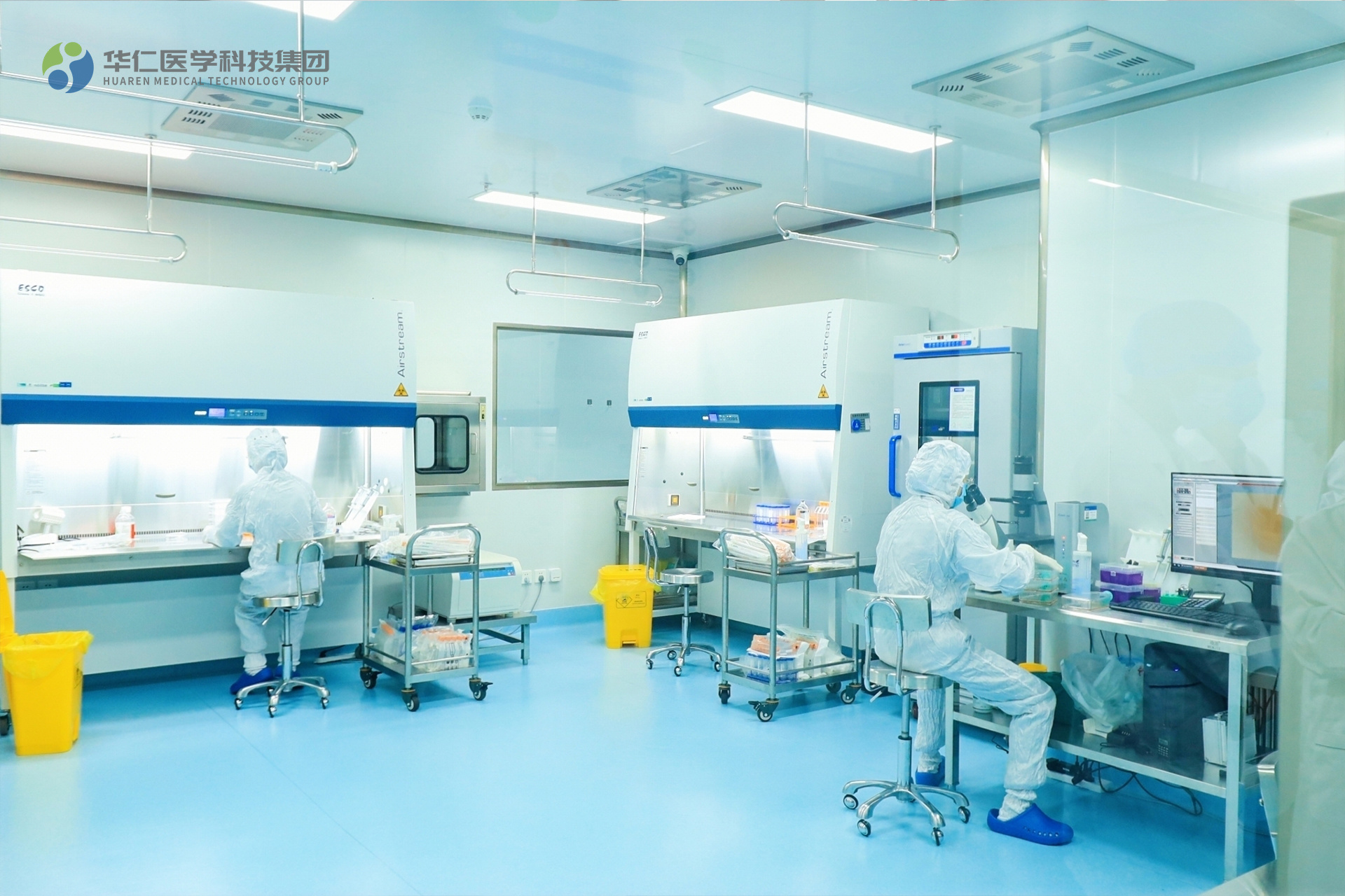
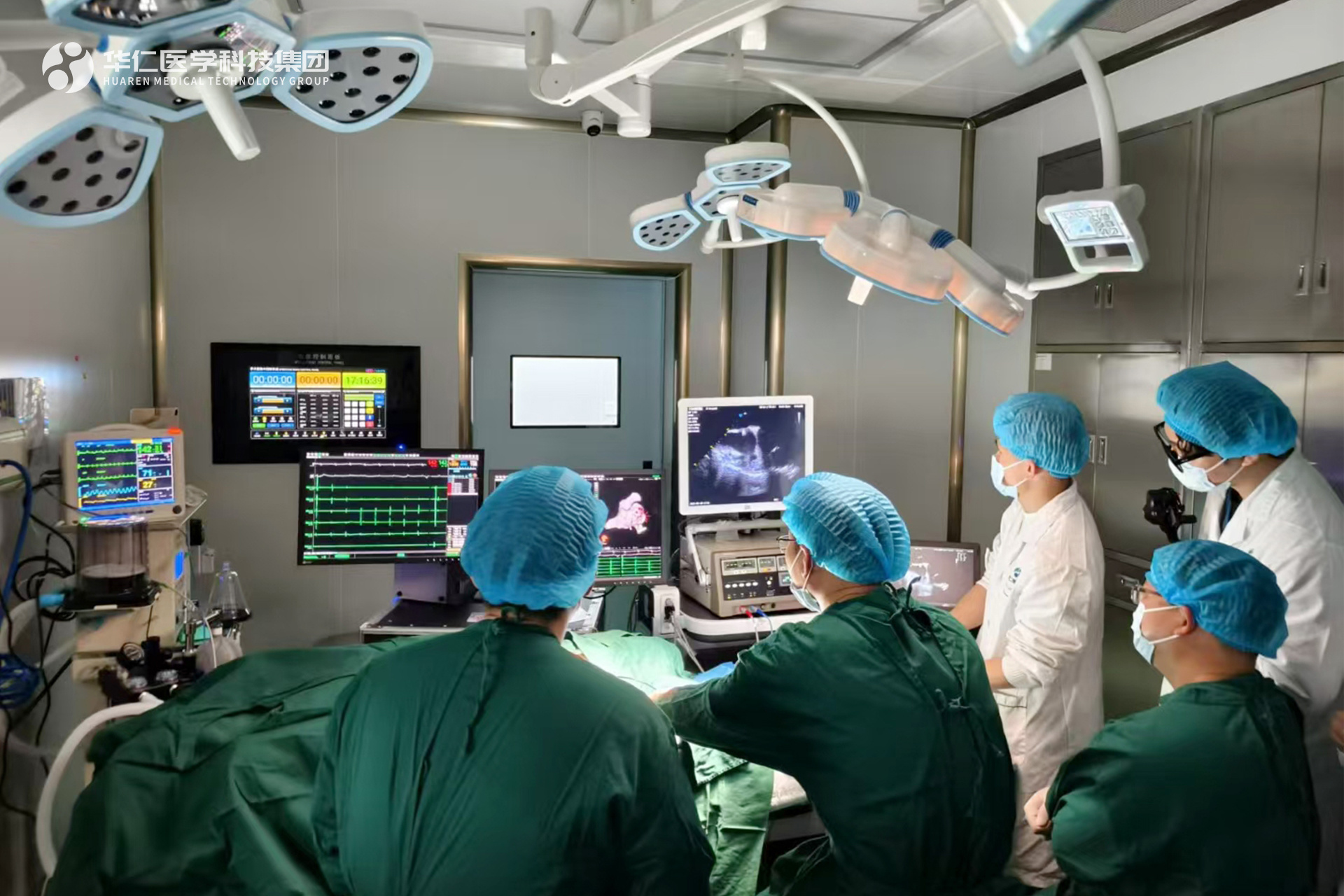 ▲On-site demonstration
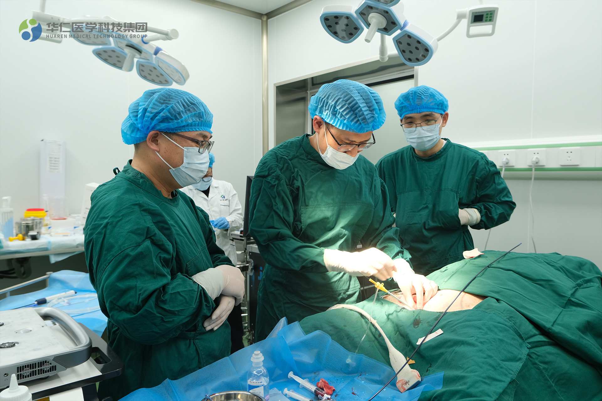
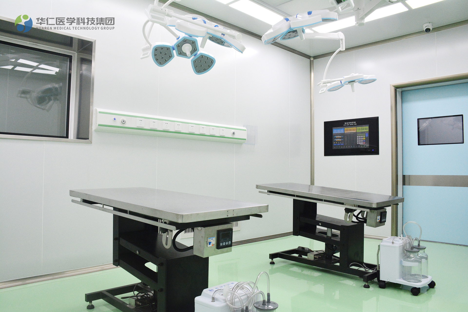
This project was conducted at the Huaren Medical Shared Laboratory-Experimental Animal Center, leveraging its advantages in advanced experimental facilities and rigorous management to provide comprehensive support for the entire experiment. The professional technical teams of Tingsheng Technology and Huaren Medical worked closely together, fully demonstrating their efficient collaboration and professional capabilities. White pigs were used as experimental animals, and a standardized operational process was implemented, achieving high-standard management from pre-operative preparation to post-operative evaluation.
The project focused on the clinical challenges of cardiac electrophysiology radiofrequency ablation, an advanced treatment method. From animal anesthesia, vascular puncture and catheter insertion, to electrophysiological mapping and radiofrequency ablation, the demonstration showcased standardized procedures and technical points.
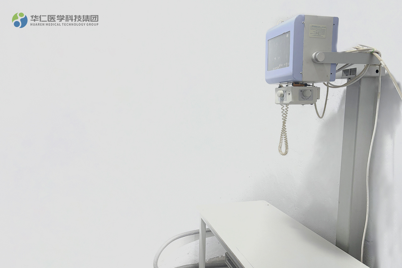
Especially in the vascular puncture and catheter insertion phase, the team used ultrasound and X-ray technology for precise positioning to ensure the accuracy and safety of the operation; during electrophysiological mapping, the three-dimensional modeling system, such as CARTO, was used to precisely analyze the electrocardiographic signals, providing a scientific basis for determining the lesion target. Finally, through rigorous post-operative monitoring and evaluation, the effectiveness and operability of the experiment were comprehensively verified.
This collaboration not only showcased high-level technical capabilities but also further highlighted the value of the Huaren Medical Technology Group's Joint Experimental Center-Shared Laboratory as a research support platform. By integrating high-quality resources and innovative service concepts, it is committed to providing research teams with "turnkey" one-stop services, helping medical research move from the laboratory to clinical application.
In the future, Huaren Medical will continue to cooperate with various research forces, promoting technological progress and scientific innovation in the medical field through multidisciplinary integration. We welcome more partners to join us in writing a new chapter in the development of life sciences.
The Huaren Medical Technology Group's Joint Experimental Center-Shared Laboratory is located at No. 9 Haihui Road, Wuxiang Investment Big Data Industrial Park Phase II, Building 3, Nanning District, China (Guangxi) Pilot Free Trade Zone. It is open for sharing nationwide and focuses on non-human primate large animal experiments and translational medical research, supporting life sciences and new drug development.
The shared laboratory is equipped with cutting-edge experimental equipment, covering a comprehensive detection and image analysis platform from the molecular and cellular to the large animal level. The experimental center has key technologies such as the construction of non-human primate animal disease models, pharmacokinetic/pharmacodynamic research, and image tracing analysis, and can provide full-process experimental support such as drug screening, mechanism of action research, disease modeling, and efficacy evaluation.
1. Non-human Primate Animal Experiment Platform
This platform focuses on large animal model research, covering pharmacokinetics, pharmacodynamics, and disease model construction. We have a standardized breeding and management system and complete animal experimental facilities. Model animals include general-purpose experimental monkeys, experimental pigs, and experimental dogs.
The service areas are wide-ranging, involving antibody drugs, cell therapy, gene therapy, and drug safety evaluation (Non-GLP) research. Featured services include the construction of various chronic and geriatric disease animal models to meet the diverse needs of scientific research and pharmaceutical development. At the same time, we deeply mine Guangxi's characteristic experimental animal resources and conduct comprehensive and professional services on Bama miniature pigs, covering breeding, disease modeling, efficacy, and toxicology research, and strive to promote the transformation and application of Guangxi's characteristic experimental animal resources, providing strong support for scientific research and industrial development.
2. Multifunctional Biochemical Analysis Platform
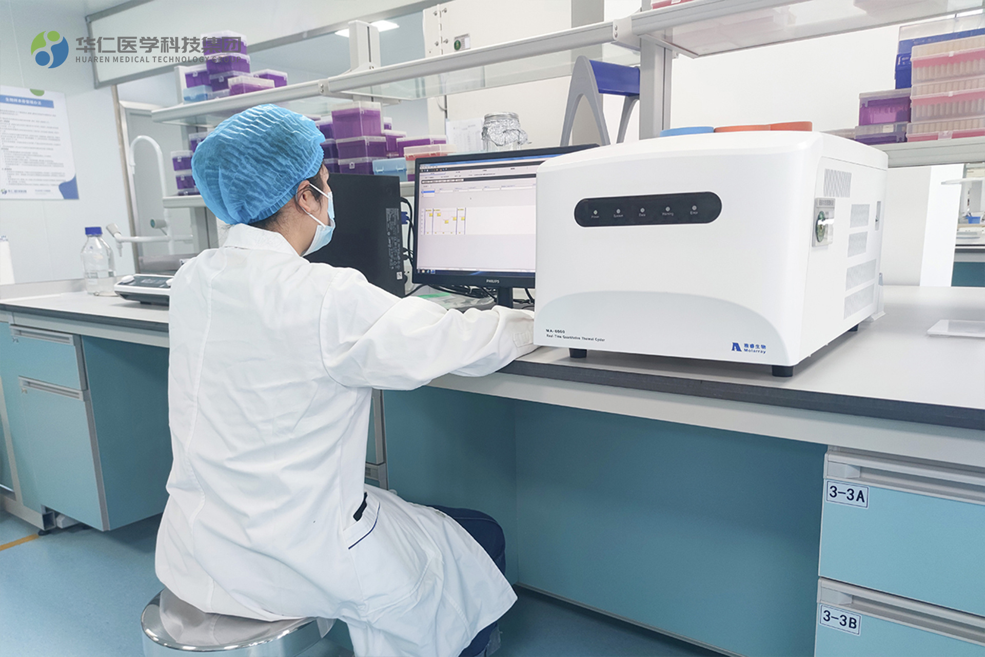
Relying on advanced equipment such as high-throughput automated enzyme-linked immunosorbent assays, protein analyzers, and flow cytometers, it can be used for high-sensitivity detection of biomarkers, cytokines, protein interactions, and signaling pathways. Suitable for drug screening and mechanism of action research in areas such as inflammation, immune regulation, metabolic diseases, and tumors.
3. Image Analysis Platform
This platform can achieve high-resolution imaging and quantitative analysis at the cellular and microtissue levels, with functions such as multi-channel fluorescence labeling, dynamic process tracking, and data analysis. Supports experimental needs such as cell viability, apoptosis, protein co-localization, and gene editing effect verification, providing accurate imaging data for drug screening, toxicity assessment, and disease research.
4. In vivo Imaging and CT Platform
It has the ability to perform in vivo imaging of small and large animals and can achieve functions such as in vivo drug tracing, disease process monitoring, and efficacy evaluation. Includes small animal in vivo optical imaging, PET-CT, MRI, large animal X-ray and CT equipment, and can conduct experimental research in areas such as cancer metastasis, brain function research, cardiovascular disease, bone disease, and tissue repair.
The experimental center is equipped with a dedicated technical and management team that provides comprehensive services such as experimental design, technical training, data analysis, and application development to ensure that researchers receive efficient and accurate experimental support.
Huaren Medical Technology Group Joint Experimental Center-Shared Laboratory, looking forward to your joining, to explore the frontier field of translational medicine together!
① Animal Anesthesia and Preoperative Preparation:
For white pig cardiac electrophysiology radiofrequency ablation experiments, the dosage of anesthetic is determined according to weight, age, and duration, such as pentobarbital sodium, which is prepared for injection according to body weight per kilogram to prevent anesthetic abnormalities. After anesthesia, fix the white pig, connect the breathing, electrocardiogram, and body temperature monitoring equipment, and place a warming pad due to thick fat, and connect the respiratory support equipment to adjust parameters for stable breathing.
② Vascular Puncture and Catheter Insertion:
Jugular or femoral vein puncture is commonly chosen for white pigs. The jugular vein is located on the side of the neck, anterior to the sternocleidomastoid muscle, while the femoral vein is located below the midpoint of the inguinal ligament. In a sterile room, the operator, wearing sterile equipment, uses a sterile needle and wire for puncture. Taking the jugular vein as an example, after disinfection, the left hand is used to fix the vein, and the right hand inserts the needle at a 30-45 degree angle. Once blood return is observed, a guidewire is inserted, followed by a dilator and sheath. A catheter is then advanced along the sheath to the heart, with X-ray or ultrasound monitoring to ensure proper placement and prevent injury.
③ Electrophysiological Mapping and Target Localization:
Using systems such as CARTO or EnSite, a mapping catheter is advanced through the vascular sheath into the cardiac chambers. Electrical signals are collected and combined with positional data to create a three-dimensional model. During the white pig's heartbeat, electrical activity is recorded, and researchers analyze the strength, frequency, and conduction time of the electrical signals. In cases of arrhythmia, the data is compared to normal data to determine the origin and conduction pathway. In atrial fibrillation experiments, the areas with disordered high-frequency electrical signals are potential targets. Operators require skilled techniques and electrophysiological knowledge to accurately locate the target.
④ Radiofrequency Ablation Implementation and Parameter Control:
Parameters are adjusted based on the characteristics of the target in the white pig's heart and its relationship to surrounding structures. For targets located in the thick ventricular wall, the energy is set to 30-50 watts, and ablation is performed for 60-120 seconds to achieve lesion destruction at 60-70℃. Near important conduction bundles, the energy is reduced to 10-20 watts, and ablation is performed for 30-60 seconds. During ablation, electrical activity and impedance are monitored, and parameters are adjusted based on feedback to preserve conduction function. Precise control is crucial.
⑤ Postoperative Monitoring and Evaluation:
After the procedure, the white pig is transferred to a monitoring room for continuous monitoring of heart rate, rhythm, respiration, and body temperature. An electrocardiogram monitor records data for 24 hours to prevent arrhythmia recurrence. Respiration is observed to check for complications, and body temperature is measured regularly to check for infection.
Here, we sincerely invite researchers from all sectors to join us, work together, and make full use of the high-quality platform of the Huaren Medical Shared Laboratory to jointly open a new chapter in medical research innovation and contribute to the development of life sciences.
SUBSCRIPTION INFORMATION
We will send you real-time information regularly!
Guangxi Huaren Medical Technology Group Co., Ltd.
Tel: 400-138-8669/ 0771-611-0603
Address: 15th Floor, Building A, Biyuan Center, Wuxiang New District, Nanning City, China (Guangxi) Pilot Free Trade Zone

Huaren Medical Technology
Huaren Medical Research
Hua Ren Cell said